
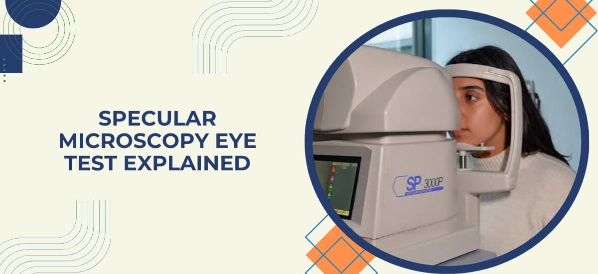
Specular microscopy is a quick, painless scan that counts and photographs the cells lining the inside of your cornea, known as the endothelium. These cells keep the cornea clear by pumping excess fluid out, and once damaged, they don't grow back. The test matters most before cataract surgery, before a corneal transplant, and whenever a condition like Fuchs' dystrophy needs tracking over time, since a low or declining cell count changes how safely certain procedures can be performed.
A specular microscopy reading taken as part of a general eye check usually just confirms things are fine. Being told your endothelial cell count is lower than expected is a different matter entirely, and it's worth understanding what that means before cataract or any other eye surgery at an Eye Hospital in Mumbai, rather than brushing past it.
“The endothelium is one of those tissues that doesn't forgive. Once a cell dies, the surrounding cells stretch to cover the gap, but the count itself never recovers. That's why I take this test seriously before any cataract or corneal procedure, a cornea that looks completely healthy on the surface can still have a cell count low enough to change the surgical plan.” — Dr. Vaishal Kenia, Chairman and Medical Director, Kenia Eye Hospital
What Is Specular Microscopy?
The corneal endothelium is a single layer of cells on the innermost surface of the cornea. Their job is to pump fluid out of the corneal tissue, keeping it clear rather than swollen and cloudy. A specular microscope photographs this layer directly and software counts the cells in the image, usually reporting the number as cells per square millimeter.
Two things get measured along with the raw count. Cell size and shape, called morphology, matter almost as much as the number itself; uniform, hexagonal cells indicate a healthy layer, while irregular or enlarged cells can signal early stress even when the total count still looks acceptable. The test takes only a few seconds per eye and needs no drops, no dilation, and no contact with the eye.
Curious what your endothelial cell count means for an upcoming procedure? Get it checked properly. Book an Appointment
Why Is Specular Microscopy Done?

This test shows up most often in the lead-up to surgery, where knowing the endothelial reserve changes how a procedure is planned.
- Before cataract surgery. Phacoemulsification puts some stress on the endothelium, so a low baseline count affects how the surgery is approached and what recovery looks like afterward.
- Before corneal transplant, both to assess the recipient's own endothelium and, for a donor cornea, to confirm the tissue meets the cell density needed for a successful graft.
- Monitoring Fuchs' endothelial dystrophy, a progressive condition where endothelial cells are gradually lost, tracked over time through repeated counts.
- Evaluating contact lens wearers, particularly long-term wearers, since extended lens use can be associated with gradual endothelial changes.
- After certain eye surgeries, to confirm the endothelium has tolerated the procedure well and isn't showing unexpected cell loss.
Patients already exploring cataract treatment can read more in our guide on the latest technology in cataract surgery, which touches on how pre-surgical testing shapes the approach used.
What Happens During a Specular Microscopy Test?
Nothing about this test requires preparation. A specular microscope is placed near the eye, not touching it, and briefly flashes a light while photographing the endothelial layer. You'll be asked to focus on a target inside the device and hold still for a moment. The whole process is over within seconds per eye, and normal activity resumes immediately afterward.
Results, including the cell count and an image of the cell pattern, are typically available the same visit, and your specialist reviews them alongside whatever procedure or condition prompted the test in the first place.
Not sure whether you need this test before an upcoming procedure? Ask during a consultation. Book an Appointment or call +91 75064 99962
Specular Microscopy Normal Range: How Results Are Interpreted
A cell count on its own only tells part of the story. Age, the reason for testing, and planned procedures all shape how a given number is read.
| Endothelial Cell Density | What It Typically Means |
|---|---|
| Above 2,000 cells/mm² | Generally considered healthy, adequate reserve for most procedures |
| 1,000 to 2,000 cells/mm² | Reduced reserve, surgery may still proceed with extra precautions |
| Below 1,000 cells/mm² | Significant risk of corneal swelling after surgery, requires careful planning |
These figures serve as general reference points rather than strict cutoffs. A count that looks borderline on paper might still be entirely workable depending on cell morphology and the specific procedure planned, which is why interpretation always involves a specialist rather than the number alone. For patients facing a corneal procedure, our guide on cornea treatment options covers what's available depending on the extent of corneal involvement.
When Should You Get a Specular Microscopy Test?

A handful of situations call for this test specifically, rather than it being part of every standard eye exam.
- Ahead of cataract surgery, particularly for patients over 60 or those with a history of eye surgery or trauma.
- Before being evaluated as a candidate for corneal transplant, either as the recipient or when assessing donor tissue.
- If Fuchs' dystrophy runs in your family or has already been diagnosed.
- As a long-term contact lens wearer due for a general eye health check.
- When your specialist flags it based on something seen during a routine exam.
For most patients, this is one of several tests bundled into a single pre-surgical workup rather than a separate visit.
Why Choose Kenia Eye Hospital for Specular Microscopy?
Reading an endothelial cell count correctly takes more than the number itself, it takes context from the rest of the eye and the procedure being planned around it. Kenia Eye Hospital brings more than 26 years of experience combining specular microscopy with corneal mapping and cataract assessment, so a borderline reading is never looked at in isolation.
Patients having this test done ahead of cataract or corneal surgery receive a clear explanation of what their results mean for the specific procedure planned, not just a number on a report. That clarity is part of what places Kenia among the best eye hospital in Mumbai choices for pre-surgical eye evaluation.
Preparing for cataract or corneal surgery? Make sure your endothelial health has been checked. Book an Appointment or call +91 75064 99962
