Keratoconus doesn't stay still. Left alone, the cornea keeps thinning, the cone keeps steepening, and vision gets progressively harder to correct. Collagen cross-linking interrupts that process. It won't reverse damage already done, but at the right stage it stops things from getting worse. For most patients that's the difference between managing the condition with lenses for decades and eventually ending up in a transplant conversation.
The technique has been in clinical use for over twenty years and the evidence is solid. What's changed is how it's done. Faster protocols, epithelium-on options, better pre-surgical mapping. The procedure itself hasn't changed much. The precision around patient selection has.
Dr. Vaishal Kenia, Chairman and Medical Director at Kenia Eye Hospital in Mumbai, says: "Cross-linking is the single most important intervention we have for keratoconus. The question is never really whether to do it, it's when. Catch it progressing and treat it early, and most of these patients never need anything more invasive."
How Does Collagen Cross-Linking Work for Keratoconus?
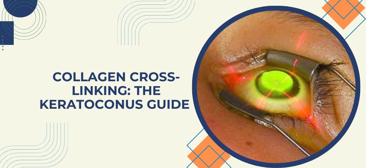
The cornea gets its strength from collagen fibres running through it in a lattice. In keratoconus those fibres weaken and the cornea starts bulging forward. Cross-linking creates new bonds between those fibres, stiffening the tissue and stopping further deformation. Here's how the procedure works step by step:
Riboflavin Drops: A form of Vitamin B2 applied repeatedly to the corneal surface over 30 minutes. In standard CXL the epithelium is removed first for deeper penetration. In transepithelial CXL a modified formula passes through the intact surface. Either way the riboflavin saturates the tissue and acts as a photosensitiser for what comes next.
UV Activation: Controlled ultraviolet A light applied for around 30 minutes. The UV activates the riboflavin and triggers new bonds between adjacent collagen fibrils. More bonds, stronger cornea, less bulging.
Accelerated vs Standard: Standard CXL uses lower UV intensity over a longer time. Accelerated CXL delivers the energy dose faster, dropping UV chair time to around ten minutes in selected protocols. The right protocol depends on the corneal findings and the surgeon's assessment.
Who Actually Needs It: Patients where serial topography confirms active progression. One scan isn't enough. Two scans over six to twelve months showing steepening or thinning confirms the disease is moving and treatment is warranted.
For a full picture of how keratoconus is diagnosed and staged, the keratoconus treatment page covers the workup in detail.
What to Expect: Recovery, Results and Limitations
Recovery is manageable for most people, but the results need to be understood clearly before going in. Here's what the process actually looks like:
First Week: Vision is blurry, sometimes worse than before the procedure. That surprises people who weren't warned. It's just the cornea healing. A bandage contact lens sits over the eye and antibiotic plus anti-inflammatory drops are used regularly. Most people are back to normal activity within seven to ten days.
What CXL Does: Stops progression. Topography at three, six, and twelve months should show a stable corneal shape. The keratoconus doesn't advance further in the majority of treated eyes. That's the goal and it's well achieved.
What CXL Doesn't Do: Restore the existing cone to a normal corneal shape. The prescription may not improve, and patients who needed rigid lenses before the procedure may still need them after. What changes is the trajectory, and lenses work far better on a cornea that isn't still shifting underneath them.
Long-term Results: Studies with ten-plus years of follow-up consistently show stabilisation holds. Repeat cross-linking is occasionally done if progression resumes but that's uncommon. Combining topo-guided PRK with CXL is an option in select cases where thickness and topography allow it.
For patients where the disease has advanced beyond what CXL can address, the cornea treatment page covers surgical options including DALK and penetrating keratoplasty.
Why Choose Kenia Eye Hospital
Kenia Eye Hospital has been in Santacruz (West), Mumbai since 1998. Over 26 years of corneal and refractive surgical work, with keratoconus and cross-linking forming a regular part of the caseload throughout. Dr. Vaishal Kenia handles the staging and timing decisions that determine whether CXL alone is sufficient or whether a combined approach makes more sense for a given patient.
Pre-surgical mapping runs through Pentacam HR and Corvis ST, assessing both corneal shape and biomechanical strength before anything is scheduled. NABH, QAI, and FEQH accredited. CGHS empanelled. Prescription keeps shifting or someone's flagged keratoconus on a scan? A corneal topography is the right starting point. Call +91 75064 99962 to book.